
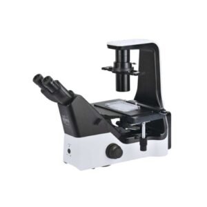


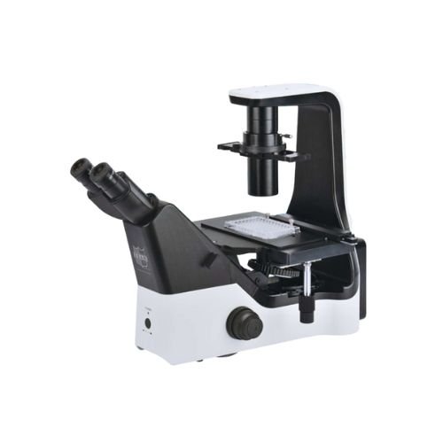
Inverted Biological Microscope – iMX50
- Make Reasonable improvement on basis of scientific research microscope.
- More suitable for laboratory observation of cells.
- Adopt long life LED light source and infinity optical system, easy to obtain high definition and high contrast wide viewing images.
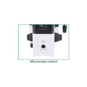
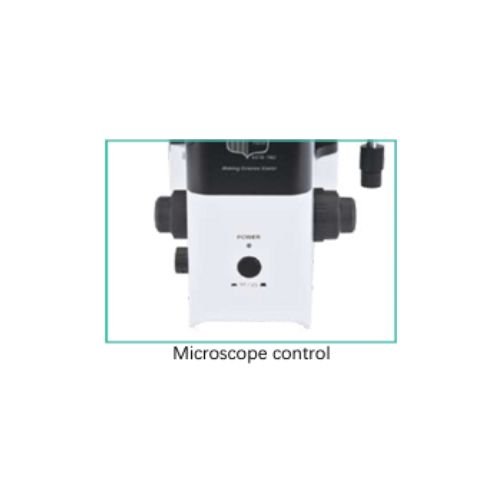
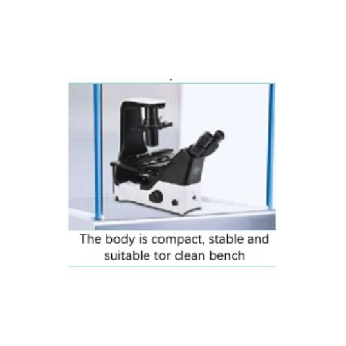
- The body is compact and stable, and the operation buttons are well arranged, the cells can be observed, sampled and processed in the super clean bench freely.
- Using 3 different color filter, it widely enlarges selectivity for dye.
- LED illumination with large intensity and even brightness provides support for high quality fluorescence observation.
- With standard camera port, QUICKLAB camera and image processing software, providing low noise, high sensitivity and resolution image.
Compare
- Optical System: NIS60 Infinite Optical System (F200).
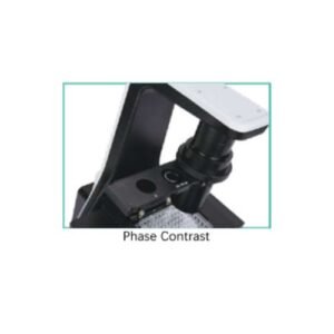
- Observation Method: Brightfield. Phase Contrast, Hoffman, phase Contrast, Emboss Contrast. Epi-Fluorescence.
- Illumination: 3W S-LED Kohler Illumination, LED illuminator, built-in Fly-eye lens, Can be configured with up to 3 different fluorescence LED units; available wavelengths:365, 405, 485, 525nm.
- Viewing Head: Seidentopf Binocular Viewing Head, Inclined at 5-35°, Interpupillary 48-75mm; Additional camera port eyepiece /port 100/0 : 0/100.
- Eyepiece (F.O.V): SW10x(22), WF15x(16), WF20x(12).
- Focusing: Coaxial coarse and fine adjustment, the function of coarse tightness adjustment, Fine Division 1 um, Fine stroke 0.2mm per rotation, Coarse stroke 37.5mm per rotation. Up 7mm, downl .5mm.
- Nosepiece: Quintuple Nosepiece.
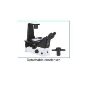
- Condenser: Condenser NA 0.3, WD 75mm, without Condenser WD 187mm.
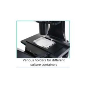
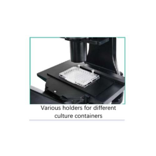
- Stage: stage: 170 (X) 250 (Y)mm Attachable Mechanical Stage: 129 (X) x 80 (Y), Accepts 5 types of micro-testplate, well clamper and stage clip.
- Holder: Petridish Holder 90mm, Slide Glass Holder for glass slides, Universal Holder for Terasaki plate holder and glass slide.
- Phase System: Condener with 4x Phase Annulus Plate 10x, 20x, 40x Universal Phase Annulus Plate.
- Hoffman Phase: 10x, 20x, 40x Hoffman Condenser, Special objective.
- Relief 3D Contrast: Condenser and Eyepiece with Emboss Contrast 10x, 20x, 40x, Universal Emboss contrast slide.
- EPI-Fluorescence Attachment: Filter cubes with noise terminator mechanism Configure with up to 3 Epi-fluorescence filter cubes, Attachable Contrast Shield.
- Dimensions: 243 (W)x587 (D)x 504 (H)mm.
- Video Adapter: 1x, 0.5x, C Mount.
- Accessories: ECO (No operator, turn off the light source automatically in 15 minutes) Heating Stage.
| GTIN |
|---|
You must be logged in to post a review.
Q & A
You may also like…
-
Microscopes
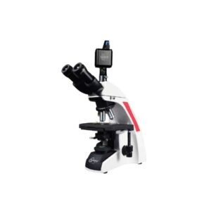
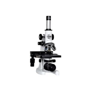
Biological Microscope – ULTIMATE MX50
Sense Series Microscope supports premium optics that delivers crisp, bright, and consistent visualization. The microscope frame well fits the hands and the location of the control knobs maximizes ergonomics to improve work efficiency. Users can quickly set a specimen with one hand while adjusting the focus and operating the stage with the other hand with minimal movement. The microscope features a camera port for digital imaging.
SKU: MX50 -
Microscopes
Ultimate Microscope with 4K Coms Camera – Scope – Cam 4K
- The Scope-Cam 4K series camera is the next-generation live view imaging-system with 4K resolution(Video) at 60 FPS.
- It comes with Sony Exmor CMOS sensor with high sensitivity, low dark current and no smear achieved through the adoption of R, G and B primary color mosaic filters.
- The camera uses a standard C-mount interface for maximum compatibility with various microscopy-systems.
- It can be used as a stand-alone recorder when used with an HDMI monitor or television, or live-streamed to a PC via Gigabit Ethernet (LAN) for image-capture and video-recording.
- Hardware 3D denoising, sharpness and tone mapping control functions greatly improve the image and video quality.
- The included Windows software offers image-development and measurement tools, as well as advanced compositing features such as image-stitching and extended-depth-of focus.
- With the ability to calibrate scales at multiple magnifications, the software can be used for multi-level inspection.
- For Mac and Linux, there is a lite version of the software which can capture video and still images, and includes limited processing features.
- The HDMI 4K series camera is intended to be used for the acquisition of digital images from the stereo microscope, biological microscope or online interactive teaching.
SKU: UTM-3.0 -
Microscopes
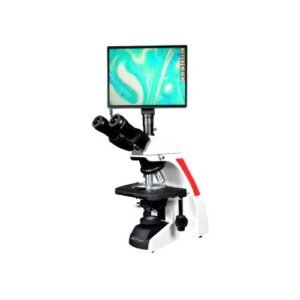
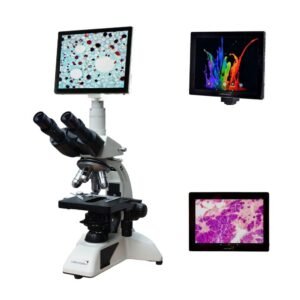
Ultimate Microscope with Digital Display- Scopepad
The Scopepad tablet with a 10.5 inch display will be supplied with 8M.P., 4K camera. Together with the Image Focus software it offers a state-of-theart solution for modern microscopy for education.
- Real-time images directly on TV, monitor or beamer.
- Stand-alone mode.
- Built-in mouse-driven software and SD memory card.
- HDMI, USB-2.0/3.0.
- GbE (Ethernet) (VC.3042).
- WiFi (VC.3042 and VC.3034).
- Compatible with Image Focus software.
SKU: Ultimate-D11


Related products
-
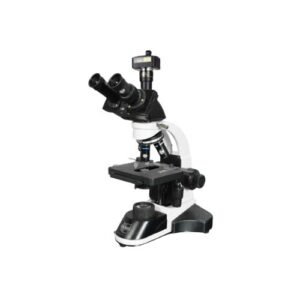
Microscopes
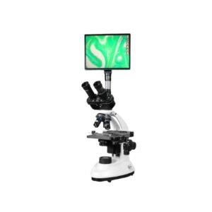
Co-Axial Microscope with Digital Display – Scopepad
The QUICKLAB Scopepad tablet with a 10.5 inch display will be supplied with 8M.P., 4K camera. Together with the Image Focus software it offers a state-of-theart solution for modern microscopy for education.
- Real-time images directly on TV, monitor or Projector.
- Stand-alone mode.
- Built-in mouse-driven software and SD memory card.
- HDMI, USB-2.0/3.0.
- GbE (Ethernet) (VC.3042).
- WiFi (VC.3042 and VC.3034).
SKU: CXL-10D -
Microscopes
Co-Axial Digital Head Teaching Microscope – Scopepad Mini
The Co-axial Microscope is also available with a 7” LCD screen, with all the features of the regular Inclined Microscope. This screen allows live viewing, saving images and videos directly to a micro SD card. The HD LCD screen can only be operated as a stand alone.
- All the Scopepad mini LCD microscopes are equipped with 7” IPS LCD screen.
- Screen resolution 1920 x 1080 @ 30 fp.
- Save images and videos directly to micro SD card.
- Image resolution upto 1844 x 1080 in .JPEG format.
- Video resolution 1280 x 720 .AVI format.
- Basic measurement function (line) with calibration.
SKU: CXL-D7 -
-
Microscopes
Co-Axial Microscope with Teaching Camera – CXL7
Digital Cameras with Next Generation CMOS Sensor
The plug and play QUICKLAB is Series Cameras produce high quality images making it a great choice for education, pathology and research applications. Available in 5,12,18,20 & 45 Megapixels All QUICKLAB is Series cameras supplied with a copy of QUICKLAB image analysis, an advanced Image processing and documentation software.Powerful, user-friendly IS Series software comes with tools for:
- Image Enhancement (hue, saturation, contrast, etc).
- 2D Measurement including calibration.
- Auto-exposure and single click white balance.
- Capture Images, High Quality Videos.
- Compatible with Windows, Mac OS and Linux.
- C/C++, C#, DirectShow, Twain Control API.
SKU: CXL-5T -
Microscopes
Trinocular Microscope – KX i2000TRI With LCD
9.7” 2nd Generation HD LED Backlit LCD Screen (1024*768)
G-sensor: Multi-point Capacitance Touch Screen
Mega Pixels: 5.0MP
Resolutions: Selectable resolution for capturing images
Image-Previewing Rate: 30fps at Full Resolution
Video Format: 720P/30fps
White Balance Setting: Manual / Automatic / Preinstall Operation
Programmable Controls: Gain, Frame Rate, Frame Size, ExposureSKU: KXi2000LCD

Reviews
There are no reviews yet